
Quality, Testing & COAs
What Does “0 Cells/mL” Mean on an Extracellular Vesicle COA?
A COA can report 0 cells/mL and 50 billion EV per 3 mL vial at the same time. Here's why cells and extracellular vesicles are not the same measurement.
When reading an extracellular vesicle Certificate of Analysis, one result can initially seem confusing:
Cell Count: 0 cells/mL
If extracellular vesicles originate from cells, how can an EV preparation contain zero cells?
And if a vial contains billions of extracellular vesicles, how can the cell count still be zero?
The answer is straightforward:
Cells and extracellular vesicles are not the same thing.
Extracellular vesicles are membrane-bound particles released by cells. They can remain in a preparation after the intact cells that produced them have been separated from it.
For HydroKarma lot KH-0007DE, KWEHEALTH's batch-specific Certificate of Analysis reports:
Cell Count: 0 cells/mL
The same COA separately reports:
EV Quantity: 50 billion EV per 3 mL vial
Those results are not contradictory. They describe two different things.
What Is a Cell Count?
For KH-0007DE, the unit is:
cells/mL
or: cells per milliliter.
A reported result of:
0 cells/mL
therefore concerns the presence of detectable cells under the conditions of the reported measurement.
It does not mean that the preparation contains zero extracellular vesicles, zero proteins, zero nanoparticles or zero dissolved material.
What Is an Extracellular Vesicle?
Extracellular vesicles are produced and released by cells, but they are not themselves intact cells.
This distinction is fundamental to EV biology.
MISEV2023, the International Society for Extracellular Vesicles' consensus guidance, describes extracellular vesicles as particles naturally released from cells that are delimited by a lipid bilayer and cannot replicate.
That last point helps illustrate the distinction.
A cell is a complex biological unit capable of carrying out cellular processes.
An extracellular vesicle is a particle released by a cell.
The core distinction
Cell ≠ extracellular vesicle
How Can EVs Exist Without Intact Cells?
Imagine cells growing in a controlled culture environment.
Those cells release extracellular vesicles into the surrounding material.
During downstream processing, the intact cells can be separated from material containing the extracellular vesicles.
The EVs don't have to remain physically attached to the cells that released them.
A simplified conceptual sequence looks like this:
- Cells
- Cells release extracellular vesicles
- EV-containing material is collected
- Intact cells and other material are separated during processing
- EV preparation undergoes further processing and characterization
This is a general educational illustration of EV processing.
It is not a description of HydroKarma's proprietary manufacturing protocol.
For a broader explanation, see How Are Exosomes Produced and Processed?.
A Real HydroKarma Example
According to KWEHEALTH's batch-specific COA for HydroKarma lot KH-0007DE:
Cell Count: 0 cells/mL
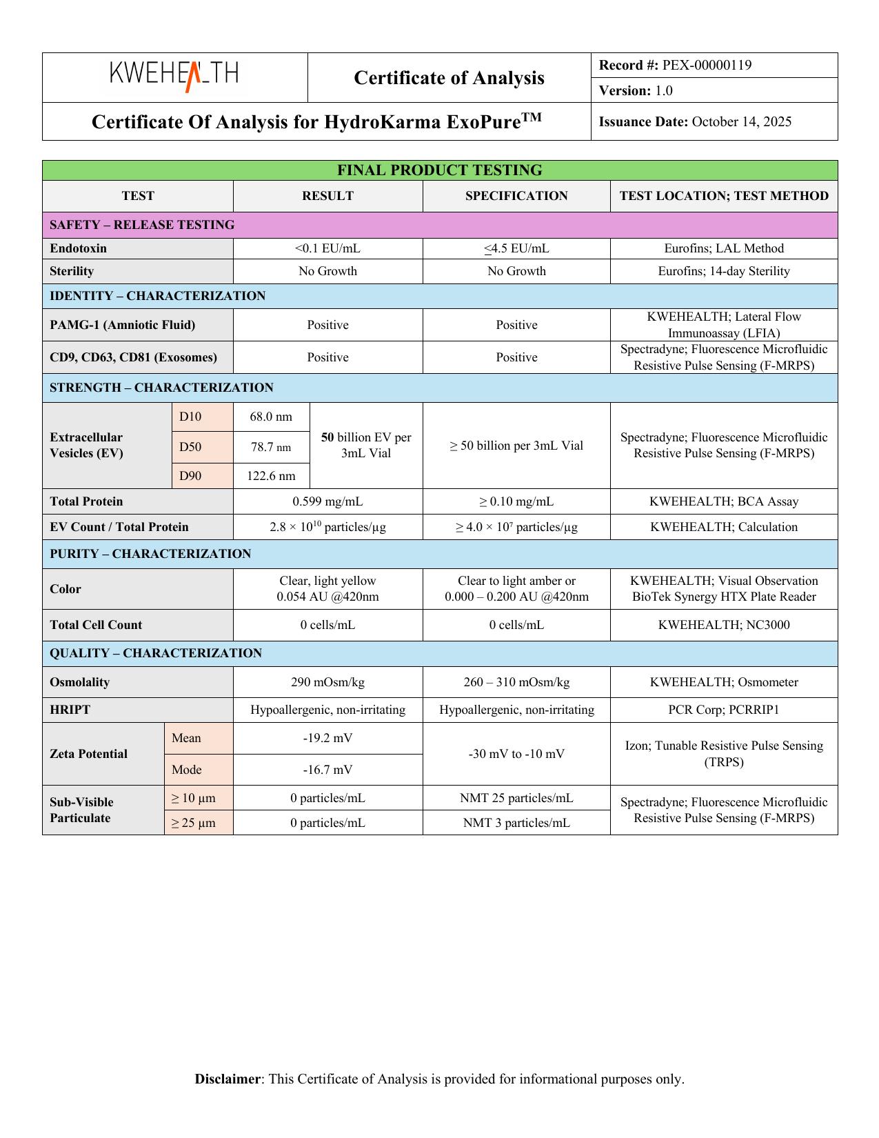
The same Final Product Testing section reports:
EV Quantity: 50 billion EV per 3 mL vial
and:
- D10: 68.0 nm
- D50: 78.7 nm
- D90: 122.6 nm
These measurements address different populations.
| Measurement | Reported Result | What It Describes |
|---|---|---|
| Cell Count | 0 cells/mL | Reported intact-cell count |
| EV Quantity | 50 billion EV per 3 mL vial | Reported EV quantity |
| D10/D50/D90 | 68.0 / 78.7 / 122.6 nm | Reported particle-size distribution |
A zero cell count does not conflict with a large EV count because EVs are not cells.
What Does “0 Cells/mL” Actually Mean?
The safest interpretation is the literal one:
KWEHEALTH reports a cell-count result of 0 cells/mL for lot KH-0007DE.
The result should remain attached to:
- the specific lot
- the reported analytical method
- the sample tested
- the measurement conditions
It should not automatically be expanded into broader claims the test does not establish.
What Method Does the HydroKarma COA Report?
Unlike several other COA rows where the analytical method isn't identified, the KH-0007DE documentation reports a method for cell count:
NC3000
This refers to the NucleoCounter NC-3000, an automated cell-analysis platform.
That distinction matters because we don't have to guess how the cell-count result was obtained—the COA actually identifies the reported method.
However, knowing the instrument name does not give us permission to invent the exact assay configuration.
Unless KWEHEALTH's documentation provides additional information, we should not assume details such as:
- staining protocol
- reagent combination
- sample preparation
- dilution
- gating criteria
- detection threshold
- replicate count
- instrument settings
The correct statement is simply:
What the COA supports
KWEHEALTH's COA reports 0 cells/mL using NC3000.
What Is the NucleoCounter NC-3000?
The NucleoCounter NC-3000 is an automated fluorescence-imaging cytometer designed for cell analysis and counting applications.
Depending on the assay and configuration, instruments of this type can use fluorescent labeling and image-based analysis to evaluate cells.
But the important distinction for this article is not the exact instrument mechanics.
It's that the cell-count measurement is fundamentally different from the EV particle measurement.
NC3000 vs. F-MRPS
This COA gives us an especially useful example of why methods matter.
KWEHEALTH reports:
Cell Count → NC3000
while the EV particle quantity and size measurements are reported using:
Spectradyne; Fluorescence Microfluidic Resistive Pulse Sensing (F-MRPS)
These methods answer different questions.
| Method | COA Measurement |
|---|---|
| NC3000 | Cell Count |
| F-MRPS | EV quantity / particle-size distribution |
We should not interchange them.
The NC3000 cell-count result does not establish the EV particle quantity.
Likewise, F-MRPS particle counting does not substitute for the reported cell-count measurement.
Why Are Cells So Much Larger Than EVs?
Cells and extracellular vesicles operate on very different size scales.
Many mammalian cells are measured in micrometers.
The EV size distribution reported for KH-0007DE is measured in nanometers.
Remember: 1 micrometer = 1,000 nanometers
The HydroKarma COA reports:
D50 = 78.7 nm
By comparison, an intact mammalian cell may be many micrometers across.
The precise size depends heavily on cell type and biological state, so there is no single universal “cell size.”
But the scale difference helps explain why an analytical method intended to count intact cells and a method intended to characterize nanoscale particles are not interchangeable.
For more on EV size, see What Does Extracellular Vesicle Size Tell Us?.
Does 0 Cells/mL Mean the Product Is 100% Pure EVs?
No.
This is probably the most important limitation to understand.
A result of 0 cells/mL does not mean:
100% of everything in the preparation is an extracellular vesicle.
Other material can exist without being an intact cell.
Depending on a preparation, that could include things such as:
- soluble proteins
- protein complexes
- nanoscale non-EV particles
- formulation components
- other noncellular material
Therefore:
Not a purity claim
0 cells/mL ≠ 100% EV purity
Cell count and EV purity are different analytical concepts.
For more on this distinction, see What Is Extracellular Vesicle Purity?.
Does 0 Cells/mL Prove Every Particle Is an EV?
No.
A cell-count measurement asks whether cells are detected according to the method.
It does not identify every nanoscale particle in the preparation.
Therefore:
- 0 cells/mL ≠ every particle is an EV
- 0 cells/mL ≠ every particle is an exosome
Particle identity requires other forms of characterization.
This is one reason the KH-0007DE COA contains additional measurements, including:
- CD9: Positive
- CD63: Positive
- CD81: Positive
Those results provide different information from the cell-count result.
See What Are CD9, CD63 and CD81?.
Does 0 Cells/mL Mean There Is No Cellular Material?
Not necessarily.
This distinction is subtle but important.
An intact cell is not the same thing as every molecule that could originate from a cell.
A preparation could have no detectable intact cells under a cell-count method while still containing biological material originating from cells.
Extracellular vesicles themselves are the obvious example: they are released by cells.
Proteins and other noncellular biological material may also be present.
Therefore:
Read it literally
0 cells/mL ≠ zero cell-derived material. The result concerns the measured cell count.
What About Cell Debris?
Cell debris is also not necessarily equivalent to an intact cell.
Depending on the analytical method, fragments of cellular material may behave differently from intact cells.
A result of 0 cells/mL should therefore not automatically be rewritten as:
“zero cellular debris.”
Unless the analytical method and manufacturer documentation specifically establish that conclusion, it goes beyond what the reported cell count tells us.
So: 0 cells/mL ≠ proof of zero cell debris
Does 0 Cells/mL Mean the Preparation Is Sterile?
No.
Cells and microorganisms are not interchangeable analytical categories.
The KH-0007DE COA separately reports:
Sterility: No Growth
That is a different test.
A cell-count instrument isn't a substitute for sterility testing.
Therefore:
- 0 cells/mL ≠ No Growth
- 0 cells/mL ≠ proof of sterility
See What Is Sterility Testing? Understanding “No Growth” on a COA.
Does 0 Cells/mL Mean There Is No Endotoxin?
No.
KWEHEALTH separately reports:
Endotoxin: <0.1 EU/mL
Endotoxin testing evaluates bacterial endotoxin.
Cell counting evaluates cells according to the cell-count method.
Therefore: 0 cells/mL ≠ zero endotoxin
See What Is Endotoxin Testing and Why Does It Matter?.
Does 0 Cells/mL Mean There Are No Large Particles?
No.
Particulate-matter testing is another separate measurement.
For KH-0007DE, KWEHEALTH reports:
- Particulate Matter ≥10 µm: 0 particles/mL
- Particulate Matter ≥25 µm: 0 particles/mL
Those results should not be inferred from the cell-count measurement.
Likewise, the particulate-matter results do not substitute for cell counting.
See What Is Particulate Matter Testing?.
Why Include Cell Count on an EV COA?
A cell-count result provides another piece of information about the finished preparation.
If the product is intended to contain extracellular vesicles rather than intact source cells, evaluating the preparation for detectable cells can help characterize whether intact cells are detected in the tested sample.
But the result must remain in context.
It does not independently tell us:
- EV concentration
- EV identity
- EV purity
- protein content
- endotoxin level
- sterility
- particulate matter
- biological activity
That's why it appears alongside other tests rather than replacing them.
Cell Count vs. Particle Count
This distinction is worth making explicit.
Cell count asks:
How many cells were detected per unit volume under the cell-count method?
EV particle count asks:
What particle quantity was reported under the EV particle-analysis method?
For KH-0007DE:
- Cell Count: 0 cells/mL — NC3000
- EV Quantity: 50 billion EV per 3 mL vial — F-MRPS
Both can be true simultaneously.
Cell Count vs. EV Purity
Cell count: looks for cells according to the analytical method.
EV purity: concerns the composition of the broader EV preparation and the extent to which measured material represents EV-associated versus other components.
Therefore, removing intact cells is not the same thing as demonstrating EV purity.
This is an important distinction when evaluating EV manufacturing and characterization.
Cell Count vs. EV Identity
Likewise: absence of detectable cells does not establish the identity of nanoscale particles.
EV identity is evaluated through a broader characterization strategy.
MISEV2023 emphasizes the importance of using complementary characterization approaches rather than relying on a single measurement to establish an EV preparation's characteristics.
That is exactly what the KH-0007DE testing panel illustrates.
How Should You Read Cell Count on a COA?
When you see a cell-count result, ask:
1. What result is actually reported?
For KH-0007DE: 0 cells/mL
2. What method is identified?
For KH-0007DE: NC3000
3. Is the result being confused with EV particle count?
Cells and EVs are different populations.
4. Is someone turning the result into a purity claim?
0 cells/mL does not establish 100% EV purity.
5. Is someone claiming there is no cell-derived material?
That conclusion doesn't follow from a zero intact-cell count.
6. Is someone interpreting it as sterility?
Cell counting and sterility testing are separate.
7. Is someone treating it as proof of exosome identity?
A zero cell count does not identify nanoscale particles.
Putting the KH-0007DE Results Together
The cell-count result becomes much more informative when interpreted alongside the rest of the Final Product Testing panel:
| Measurement | Reported Result | What It Helps Describe |
|---|---|---|
| EV Quantity | 50 billion EV / 3 mL | Particle quantity |
| D10/D50/D90 | 68.0 / 78.7 / 122.6 nm | Particle-size distribution |
| CD9/CD63/CD81 | Positive | Selected EV-associated markers |
| Total Protein | 0.599 mg/mL | Bulk protein concentration |
| EV Count / Total Protein | 2.8 × 10¹⁰ particles/µg | Particle-to-protein relationship |
| Cell Count | 0 cells/mL | Reported cell count |
| Zeta Potential | −19.2 / −16.7 mV | Electrokinetic characteristics |
| Endotoxin | <0.1 EU/mL | Bacterial endotoxin |
| Sterility | No Growth | Microbial-growth testing |
| Osmolality | 290 mOsm/kg | Osmotic concentration |
| Particulate Matter ≥10 µm | 0 particles/mL | Larger-particle count at specified threshold |
| Particulate Matter ≥25 µm | 0 particles/mL | Larger-particle count at specified threshold |
Again, no single measurement is a universal quality score.
Each answers a narrower analytical question.
For the complete framework, see How to Read an Exosome Certificate of Analysis (COA).
The Bottom Line
For HydroKarma lot KH-0007DE, KWEHEALTH reports:
Cell Count: 0 cells/mL
using: NC3000
The same COA reports:
50 billion EV per 3 mL vial
using F-MRPS for the reported EV quantity/size characterization.
Those results are not contradictory because: cells and extracellular vesicles are different things.
A reported cell count of zero should not automatically be interpreted as:
- zero EVs
- zero cell-derived material
- zero cellular debris
- 100% EV purity
- every particle being an EV
- every particle being an exosome
- sterility
- zero endotoxin
- zero particulate matter
Instead, read the result for what it actually tells you:
The literal reading
KWEHEALTH reports 0 cells/mL under the cell-count measurement for lot KH-0007DE. That is one piece of the larger characterization picture.
Original Source / References
- Welsh et al. — Minimal information for studies of extracellular vesicles (MISEV2023): From basic to advanced approaches. Journal of Extracellular Vesicles, 2024. MISEV2023 provides consensus guidance on extracellular-vesicle terminology, separation and characterization. DOI: 10.1002/jev2.12404 (PMID 38326288)
- ChemoMetec — NucleoCounter NC-3000. Manufacturer technical documentation describing the NC-3000 automated fluorescence-imaging cytometer and its cell-analysis capabilities. This source should be used only for general information about the instrument, not as evidence for HydroKarma's specific assay configuration.
- KWEHEALTH/HydroKarma batch-specific Certificate of Analysis — Lot KH-0007DE. The 0 cells/mL, NC3000, EV quantity and other HydroKarma-specific values discussed here come from manufacturer-supplied batch documentation and should not be attributed to MISEV2023 or ChemoMetec.
Continue Learning
What Is Extracellular Vesicle Purity? How EV Purity Is Evaluated
Extracellular vesicle purity is a multidimensional characterization question, not a single number on a Certificate of Analysis. Here's how particle counts, protein measurements, markers and particle-to-protein ratios each contribute — and what none of them establish alone.
What Are CD9, CD63 and CD81? Understanding Common Extracellular Vesicle Markers
CD9, CD63 and CD81 are tetraspanin proteins commonly evaluated when characterizing extracellular vesicles. Their detection is a core part of how EV preparations are characterized, and KWEHEALTH/HydroKarma uses these three markers as the basis of its reported exosome count.
How to Read an Exosome Certificate of Analysis (COA)
A plain-English walkthrough of an extracellular-vesicle Certificate of Analysis: lot numbers, analytical methods, particle concentration, particle size, characterization, specifications and results.
Understanding Extracellular Vesicle Characterization and Testing
Characterization is how laboratories measure and describe different properties of an extracellular-vesicle preparation — particle count, particle size, EV-associated markers, total protein and zeta potential — and why no single test describes everything.