
Quality, Testing & COAs
What Are CD9, CD63 and CD81? Understanding Common Extracellular Vesicle Markers
CD9, CD63 and CD81 are tetraspanin proteins commonly evaluated when characterizing extracellular vesicles. Their detection is a core part of how EV preparations are characterized, and KWEHEALTH/HydroKarma uses these three markers as the basis of its reported exosome count.
If you've looked at an extracellular vesicle Certificate of Analysis or read scientific literature about exosomes, you've probably encountered three names repeatedly:
- CD9
- CD63
- CD81
On the KWEHEALTH/HydroKarma Certificate of Analysis we've been examining throughout this Learn series, all three are reported as:
Positive
But what exactly are CD9, CD63 and CD81?
Why do researchers look for them?
And how are they used when a manufacturer reports an exosome count?
CD9, CD63 and CD81 are tetraspanin proteins found in the membranes of extracellular vesicles, and they are among the markers widely used to characterize EV preparations. Detecting them is meaningful evidence, and it is read alongside the rest of a preparation’s characterization rather than on its own.
Understanding that distinction makes the results on an EV Certificate of Analysis much more meaningful.
What Are CD9, CD63 and CD81?
CD9, CD63 and CD81 belong to a family of proteins called tetraspanins.
Tetraspanins are membrane proteins found in many types of cells and participate in the organization of cell membranes and various cellular processes.
Their name provides a clue to their structure.
CD9, CD63 and CD81 are among the tetraspanins frequently studied in extracellular-vesicle research.
Because these proteins can be enriched or detected in EV preparations, researchers often use them as part of a panel of markers when characterizing extracellular vesicles.
Why Are Tetraspanins Associated With Extracellular Vesicles?
Extracellular vesicles are membrane-bound particles released by cells.
Because EVs are surrounded by a lipid membrane, proteins associated with cellular membranes can also be present in EV membranes.
CD9, CD63 and CD81 are among the membrane-associated proteins that have become widely used in EV research.
You will often see them discussed in scientific papers, EV characterization experiments and product testing documentation.
But there's an important point:
They are not exclusive to extracellular vesicles.
These proteins also occur in cellular membranes and biological systems outside the specific EV population being studied.
That's one reason modern EV characterization doesn't generally rely on the detection of a single tetraspanin alone.
Are CD9, CD63 and CD81 “Exosome Markers”?
You'll frequently hear CD9, CD63 and CD81 called exosome markers.
That phrase can be useful conversational shorthand, but scientifically it needs some qualification.
As we discussed in Exosomes vs. Extracellular Vesicles: What's the Difference?, the term exosome technically refers to an EV associated with a particular intracellular biogenesis pathway.
Determining that a particle originated through that pathway isn't always possible simply by examining an isolated preparation.
For that reason, the International Society for Extracellular Vesicles' MISEV2023 guidance encourages careful terminology and the use of multiple approaches when characterizing EVs.
Detecting CD9, CD63 or CD81 is strong evidence that a preparation contains extracellular vesicles.
Confirming the endosomal origin that defines an exosome in the strict scientific sense requires additional evidence.
A more precise description is:
CD9, CD63 and CD81 are commonly evaluated EV-associated tetraspanins.
Why Test More Than One Marker?
Biological populations are complicated.
Not every extracellular vesicle necessarily carries exactly the same proteins or carries them at the same abundance.
EV populations can be heterogeneous, meaning they can contain particles with different characteristics.
CD9, CD63 and CD81 also aren't necessarily distributed identically among every EV subtype or biological source.
Looking at multiple markers therefore provides more information than asking whether one particular protein can be detected.
Instead of:
“Is CD63 present?”
a characterization strategy can ask:
“What combination of EV-associated proteins is detected, and what other evidence do we have about the preparation?”
That broader approach is consistent with the characterization principles discussed in Understanding Extracellular Vesicle Characterization and Testing.
What Does a “Positive” Result Mean?
Suppose a Certificate of Analysis reports:
| Marker | Result |
|---|---|
| CD9 | Positive |
| CD63 | Positive |
| CD81 | Positive |
At the simplest level, this means the manufacturer's reported testing detected the respective markers according to the method and criteria used for that test.
It does not automatically tell you:
- how much CD9 is present
- how much CD63 is present
- how much CD81 is present
- what percentage of particles carry each marker
- whether every particle carries all three markers
- whether every detected particle is an exosome
- the biological activity of the preparation
Those would require additional information or different measurements.
This distinction between a qualitative result and a quantitative result is important when reading laboratory documentation.
So when you see Positive, don't mentally translate that into:
“100% of particles contain this marker.”
That's not what the word tells you.
A Real Example From a HydroKarma COA
The KWEHEALTH/HydroKarma Certificate of Analysis for lot KH-0007DE reports:
| Characterization | Result |
|---|---|
| CD9 | Positive |
| CD63 | Positive |
| CD81 | Positive |
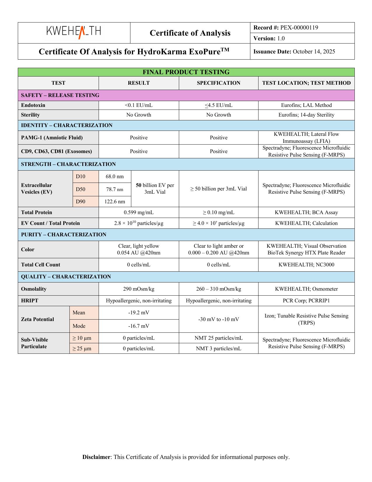
According to KWEHEALTH's batch-specific Certificate of Analysis, these are the reported marker results for that particular lot.
They should be interpreted as lot-specific manufacturer-reported characterization results.
They aren't universal specifications for every EV preparation, and they shouldn't be presented as findings from the independent scientific papers referenced in this article.
The COA provides other characterization information as well, including particle quantity and particle-size measurements.
How KWEHEALTH/HydroKarma Uses These Markers in Its Reported Exosome Count
KWEHEALTH/HydroKarma states that extracellular vesicles carrying at least one of the tetraspanins CD9, CD63 or CD81 are included in its reported exosome count.
On that basis, according to KWEHEALTH, HydroKarma's 50-billion designation means 50 billion exosomes per vial, in addition to other extracellular vesicles naturally present in the product.
This is the manufacturer’s stated counting basis for its own product. It is not a universal scientific definition of an exosome, and other manufacturers may count differently.
That's useful because EV characterization becomes much more informative when several different questions are addressed rather than relying on a single measurement.
You can see how these different measurements fit together in How to Read an Exosome Certificate of Analysis (COA).
Markers Answer a Different Question Than Particle Count
This distinction is especially useful.
A particle-count measurement asks something like:
How many particles were detected within the measurement conditions?
Marker testing asks a different question:
Were particular proteins associated with the preparation detected?
Those aren't interchangeable measurements.
Imagine a hypothetical analysis reports:
40 billion particles
and:
- CD9: Positive
- CD63: Positive
- CD81: Positive
The particle measurement doesn't automatically tell you which proteins are associated with those particles.
Likewise, positive marker results don't independently tell you that there are 40 billion particles.
The two types of testing provide different pieces of characterization information.
That's why we spent an entire article discussing Understanding Exosome Concentration and Particle Count separately.
Particle Size Answers Yet Another Question
Particle-size measurements provide another layer of information.
On HydroKarma lot KH-0007DE, KWEHEALTH reports:
- D10: 68.0 nm
- D50: 78.7 nm
- D90: 122.6 nm
using Fluorescence Microfluidic Resistive Pulse Sensing (F-MRPS) for the reported EV quantity/size analysis.
Those measurements describe the reported particle-size distribution.
They don't replace marker testing.
Likewise, marker testing doesn't replace particle-size analysis.
We can think about the information this way:
| Test Type | Basic Question |
|---|---|
| Particle quantity | How many particles were detected? |
| Particle-size distribution | What sizes were measured? |
| EV-associated marker testing | Were particular EV-associated proteins detected? |
| Other quality tests | What additional characteristics or quality attributes were measured? |
No single row answers all the others.
Why Characterization Uses More Than One Marker
Imagine a preparation where only CD63 was tested.
A single marker gives a narrow view, for several reasons.
First, CD63 isn't exclusive to exosomes.
Second, biological preparations can contain multiple types of particles and molecular components.
Third, extracellular-vesicle populations themselves can be heterogeneous.
And fourth, the term exosome describes biological origin—not simply particle size or the presence of one surface protein.
This is why MISEV guidance emphasizes multiple complementary characterization approaches rather than treating one marker as definitive proof.
What Does MISEV2023 Recommend?
MISEV2023 provides extensive recommendations for extracellular-vesicle research and characterization.
One important principle is that EV characterization should evaluate multiple categories of components and potential contaminants rather than depending on a single supposedly universal marker.
The guidelines discuss characterization of proteins associated with EVs, including membrane-associated proteins, alongside other analytical approaches.
CD9, CD63 and CD81 are widely used examples of EV-associated transmembrane proteins.
But the larger lesson from MISEV is more important than memorizing three protein names:
Strong EV characterization comes from converging evidence.
That means asking multiple questions about a preparation.
What particles are present?
What sizes are they?
What proteins are associated with the preparation?
Are potential non-EV components being considered?
What methods were used?
And what exactly do those methods allow us to conclude?
Are All Three Markers Found on Every Extracellular Vesicle?
No.
It's easy to imagine a textbook EV with CD9, CD63 and CD81 all sitting neatly on its membrane.
Real biological populations are more complicated.
Individual extracellular vesicles can differ in their molecular composition.
The relative abundance of particular tetraspanins can also vary depending on factors such as:
- cell or tissue source
- EV population
- biological state
- isolation or enrichment approach
- analytical method
Research has demonstrated heterogeneity among EV populations and differences in tetraspanin expression.
Therefore:
CD9-positive, CD63-positive and CD81-positive does not mean every individual vesicle necessarily carries all three proteins.
The reported result concerns what was detected in the tested preparation using the reported testing approach.
Why Are These Markers Useful Then?
If they're not exclusive to exosomes and aren't present identically on every EV, you might reasonably ask:
Why test for them at all?
Because scientific characterization rarely depends on one perfect marker.
Instead, researchers combine multiple pieces of evidence.
Detection of commonly EV-associated proteins can contribute to the overall characterization profile.
Combine that with information such as:
- particle concentration
- particle-size distribution
- additional protein characterization
- non-EV component assessment
- analytical methods
- purity-related measurements
- lot-specific quality testing
and you get a much more informative picture than any one test provides alone.
This is the same principle we've returned to throughout the Learn library:
Don't ask one measurement to tell you more than it actually measures.
Marker Testing and Purity Are Not the Same Thing
Another distinction worth understanding is the difference between marker detection and purity.
Finding CD9, CD63 and CD81 doesn't automatically tell you how much non-EV material is present in a preparation.
Biological samples may contain proteins, lipoproteins and other particles or components alongside extracellular vesicles depending on the source material and processing approach.
That's why broader characterization can include measurements intended to provide additional information about the preparation.
Marker testing is useful.
But:
EV-associated markers ≠ complete purity analysis
Just as:
Particle count ≠ EV identity
and:
Particle size ≠ exosome identity
Each measurement needs to be interpreted within its proper scope.
How Does This Relate to the COA?
A Certificate of Analysis becomes much easier to understand once you stop looking for a single number or result that supposedly proves everything.
Instead, read it as a collection of questions.
For example:
CD9 / CD63 / CD81
Were commonly evaluated EV-associated proteins detected?
EV quantity
What particle quantity does the manufacturer report?
Particle-size distribution
What size range or distribution was measured?
Protein concentration
What protein concentration was reported?
Sterility
What was the reported microbiological result for the lot?
Each test contributes information about a different aspect of the product.
Our How to Read an Exosome Certificate of Analysis (COA) guide walks through this approach in more detail.
Five Questions to Ask When You See CD9, CD63 and CD81
When you encounter these markers on an EV report or COA, ask:
1. Which markers were tested?
Don't assume that one marker represents an entire characterization strategy.
2. What was the testing method?
A result is more meaningful when you understand how it was generated.
3. Is the result qualitative or quantitative?
“Positive” tells you something different from a measured concentration or percentage.
4. What other characterization data are provided?
Look at marker information alongside particle quantity, size distribution and other relevant measurements.
5. Is the result lot-specific?
A batch-specific COA describes the tested lot—not every EV preparation everywhere.
Those five questions will tell you far more than simply looking for the words CD9 positive.
The Bigger Picture
CD9, CD63 and CD81 are useful because they're part of a larger characterization framework.
Think of EV characterization as assembling a puzzle.
One piece might describe particle quantity.
Another describes particle size.
Another examines EV-associated proteins.
Another considers potential non-EV material.
Another documents manufacturing or quality-control results.
One puzzle piece alone doesn't show you the entire picture.
But when several independent measurements point in compatible directions, you gain a more informative description of the preparation.
That's the purpose of characterization.
The Bottom Line
CD9, CD63 and CD81 are tetraspanin membrane proteins commonly evaluated in extracellular-vesicle research and characterization.
Their detection is meaningful evidence that a preparation contains extracellular vesicles.
A qualitative positive result tells you the marker was detected in the tested lot. It does not, on its own, tell you:
- every particle carries that marker
- every particle carries all three markers
- every particle is an exosome
- the preparation is completely pure
- a particular particle concentration has been established
- biological activity has been demonstrated
Instead, marker testing should be interpreted alongside other characterization measurements.
For HydroKarma lot KH-0007DE, KWEHEALTH reports:
- CD9: Positive
- CD63: Positive
- CD81: Positive
Those results become much more useful when considered together with the lot's particle quantity, particle-size distribution, other characterization measurements, and batch-specific quality testing.
That's the central lesson:
Good EV characterization comes from several complementary measurements read together — each interpreted within what it actually measures.
Original Source / References
- Welsh et al. — Minimal information for studies of extracellular vesicles (MISEV2023): From basic to advanced approaches. Journal of Extracellular Vesicles, 2024. DOI: 10.1002/jev2.12404, PMID: 38326288. MISEV2023 provides current consensus recommendations concerning EV terminology, characterization, marker categories and complementary analytical approaches.
- Kowal et al. — Proteomic comparison defines novel markers to characterize heterogeneous populations of extracellular vesicle subtypes. Proceedings of the National Academy of Sciences, 2016. DOI: 10.1073/pnas.1521230113, PMID: 26858453. This study examined protein composition across EV subpopulations and helps illustrate why extracellular vesicles are heterogeneous and why individual markers shouldn't be treated as universal identifiers.
- Mathieu et al. — Specificities of secretion and uptake of exosomes and other extracellular vesicles for cell-to-cell communication. Nature Cell Biology, 2019. DOI: 10.1038/s41556-018-0250-9, PMID: 30602770. This review discusses EV biology, heterogeneity, biogenesis and commonly studied proteins including tetraspanins.
- KWEHEALTH/HydroKarma batch-specific Certificate of Analysis — Lot KH-0007DE. HydroKarma-specific CD9, CD63 and CD81 results and other lot-specific measurements discussed in this article come from KWEHEALTH's supplied Certificate of Analysis and are not findings of the independent scientific publications above.
Continue Learning
What Is Extracellular Vesicle Purity? How EV Purity Is Evaluated
Extracellular vesicle purity is a multidimensional characterization question, not a single number on a Certificate of Analysis. Here's how particle counts, protein measurements, markers and particle-to-protein ratios each contribute — and what none of them establish alone.
Understanding Extracellular Vesicle Characterization and Testing
Characterization is how laboratories measure and describe different properties of an extracellular-vesicle preparation — particle count, particle size, EV-associated markers, total protein and zeta potential — and why no single test describes everything.
How to Read an Exosome Certificate of Analysis (COA)
A plain-English walkthrough of an extracellular-vesicle Certificate of Analysis: lot numbers, analytical methods, particle concentration, particle size, characterization, specifications and results.