
Manufacturing & cGMP
What Is Extracellular Vesicle Isolation and Purification?
A plain-English guide to the processes used to separate and enrich extracellular vesicles from complex biological material, the tradeoffs each approach involves, and why characterization is a separate question from processing.
Extracellular vesicles exist in complex biological fluids alongside proteins, cells, lipoproteins, cellular debris and many other biological components.
So how do researchers get from a complex starting material to a preparation enriched in extracellular vesicles?
That is where extracellular vesicle isolation and purification come in.
These terms describe processes used to separate or enrich extracellular vesicles from other components of a biological sample. Researchers have developed numerous approaches, including centrifugation, filtration, size-exclusion chromatography, density-based separation, precipitation and affinity-based techniques.
But there is an important concept to understand from the beginning:
There is no single universally perfect EV isolation method.
Different methods separate materials according to different physical or biological properties. Each approach can involve tradeoffs involving EV recovery, purity, processing time, scalability and the types of non-EV material that may remain.
Understanding those tradeoffs helps explain why EV characterization is so important after processing.
What Does Extracellular Vesicle Isolation Mean?
Extracellular vesicles are released into biological environments containing many other components.
Researchers therefore need ways to separate EVs from at least some of the surrounding material before studying them.
The word isolation can sometimes give the impression that the resulting material consists exclusively of extracellular vesicles.
That isn't necessarily what it means.
Depending on the method and starting material, an isolated or enriched EV preparation can still contain non-EV components.
This is one reason the scientific literature increasingly emphasizes describing what method was used and what was characterized, rather than assuming that the word isolated automatically means completely pure.
What Does Purification Mean?
Isolation and purification are closely related terms.
Purification generally aims to separate EV-associated material from unwanted or non-EV material.
But as we discussed in What Is Extracellular Vesicle Purity? How EV Purity Is Evaluated, EV purity isn't usually represented by one universally definitive measurement.
A preparation still needs to be characterized.
That creates a useful distinction:
- Isolation/purification = how the preparation is processed
- Characterization = how the resulting preparation is evaluated
Those concepts work together, but they aren't the same thing.
Why Is EV Isolation Challenging?
The starting materials used in EV research can be biologically complex.
Depending on the source, they may contain:
- extracellular vesicles
- soluble proteins
- protein aggregates
- lipoproteins
- cells
- cellular debris
- membrane fragments
- nucleic acids
- other biological particles and molecules
Some of these materials can share physical characteristics with extracellular vesicles.
For example, certain non-EV particles can overlap with EVs in size or density.
That creates a fundamental challenge:
A separation method targeting one physical characteristic may also capture other materials sharing that characteristic.
This is one reason researchers often use multiple processing or characterization approaches.
How Can Extracellular Vesicles Be Separated?
EV isolation methods exploit differences in physical or biochemical characteristics.
Different techniques might separate materials according to:
| Separation Principle | What It Uses |
|---|---|
| Size | Differences in particle dimensions |
| Density | Differences in buoyant density |
| Sedimentation | Differences in how materials respond to centrifugal force |
| Solubility | Differences in precipitation behavior |
| Molecular interaction | Binding to selected surface molecules |
| Filtration | Passage or retention based largely on size |
Some workflows use one approach.
Others combine several.
There isn't necessarily one method that is best for every biological source, research question or downstream application.
Differential Centrifugation
One of the best-known approaches in EV research is differential centrifugation.
Centrifugation applies force to a sample by spinning it at high speed.
Different components sediment differently depending on characteristics including size, density and the centrifugal conditions used.
A simplified research workflow might use progressively stronger centrifugation steps to remove:
cells → larger debris → larger particles → smaller EV-associated particles
Very high centrifugal forces have historically been used to pellet small extracellular vesicles.
What Is Ultracentrifugation?
Ultracentrifugation uses equipment capable of generating extremely high centrifugal forces.
It has been widely used in EV research and historically became one of the most recognizable EV isolation approaches.
However, ultracentrifugation isn't synonymous with EV purification.
Depending on the sample and protocol, other material can sediment along with EVs.
High centrifugal forces can also affect recovery and characteristics of the resulting preparation.
So:
Ultracentrifugation ≠ automatic EV purity
It is one separation technique among several.
Density-Gradient Separation
Density-based approaches attempt to separate particles according to their buoyant density.
A sample can be placed into a density gradient created using materials such as iodixanol or sucrose.
During centrifugation, different components migrate according to their physical properties.
Density-gradient methods can help separate EV-associated material from certain co-isolated components.
However, these approaches can require substantial processing time and specialized equipment.
As with other techniques, the effectiveness depends on the biological material, protocol and objective.
Size-Exclusion Chromatography
Size-exclusion chromatography, commonly abbreviated SEC, separates materials largely according to size as they travel through a column containing porous material.
Larger particles generally move through the column differently from smaller molecules that can enter more of the pores.
In EV workflows, SEC can help separate extracellular-vesicle-sized particles from some smaller soluble proteins and other molecules.
SEC has become a commonly discussed EV separation method because it can provide useful separation while avoiding some of the very high forces associated with ultracentrifugation.
But SEC also has limitations.
Particles or components with overlapping size characteristics may not be completely separated.
Again:
Size separation ≠ guaranteed particle identity
Filtration and Ultrafiltration
Filtration uses membranes or filters to separate material according to characteristics that include particle or molecular size.
In a simple example, a filter might allow smaller components to pass while retaining larger components.
Ultrafiltration uses membranes with defined molecular-weight or size-related retention characteristics and can be used to concentrate EV-containing samples.
Filtration can also be incorporated into multistep workflows.
However, membrane interactions, filter characteristics, pressure and other processing variables can influence recovery.
Filtration therefore isn't simply a matter of choosing a pore size and assuming everything retained is an extracellular vesicle.
Tangential Flow Filtration
A related technique is tangential flow filtration, often abbreviated TFF.
In conventional dead-end filtration, material flows primarily toward the filter.
In TFF, the sample flows across the surface of the membrane while part of the fluid passes through it.
This configuration can be useful for processing larger volumes and concentrating biological materials.
TFF has attracted considerable interest in EV processing because of its potential scalability.
But like other techniques, TFF separates according to physical characteristics rather than magically identifying individual extracellular vesicles.
The resulting preparation still requires characterization.
Precipitation-Based Methods
Another approach involves altering solution conditions so that extracellular-vesicle-associated material precipitates and can be collected.
Commercial precipitation reagents have made this type of EV isolation relatively convenient for some laboratory workflows.
The advantage can be simplicity and recovery.
The tradeoff is that precipitation can also collect non-EV material, including proteins and other biological components.
This illustrates one of the recurring themes in EV processing:
Recovering more material and obtaining a more selectively purified preparation aren't necessarily the same objective.
Affinity-Based Isolation
Affinity methods take a different approach.
Instead of separating particles primarily by size or density, they use interactions with selected molecules.
For example, a capture system might target proteins associated with particular extracellular-vesicle populations.
This can allow researchers to enrich specific EV-associated populations.
However, affinity-based approaches introduce their own considerations.
The captured population depends on the selected target.
If an EV doesn't express the targeted molecule—or expresses it differently—it may not be captured.
That connects directly to our discussion in What Are CD9, CD63 and CD81? Understanding Common Extracellular Vesicle Markers.
Extracellular-vesicle populations are heterogeneous.
Selecting one marker can therefore select a particular subset rather than necessarily representing every EV present in the original material.
Can Multiple Isolation Methods Be Combined?
Yes.
EV workflows frequently combine techniques because different methods address different separation challenges.
For example, a research workflow might involve:
removal of cells → removal of debris → concentration → size-based separation → characterization
Another might combine filtration with chromatography.
The exact workflow depends on factors such as:
- starting material
- sample volume
- desired EV population
- required recovery
- purity objectives
- downstream analysis
- available equipment
- processing scale
This is why asking:
“What is the best EV isolation method?”
doesn't have one universal answer.
A better question is:
“Which processing strategy is appropriate for the starting material and intended objective, and how was the resulting preparation characterized?”
Yield and Purity Can Involve a Tradeoff
Imagine a processing method recovers nearly every particle from a starting sample.
That sounds ideal.
But what if it also recovers large amounts of non-EV material?
Now imagine another method produces a much more selectively enriched preparation but loses some EV-associated particles during processing.
Which is better?
It depends on the objective.
This illustrates a common relationship between yield and purity.
A process optimized for maximum recovery isn't necessarily optimized for maximum separation from non-target material.
Likewise, highly selective purification can sometimes reduce overall recovery.
This tradeoff is one reason particle count alone shouldn't be used to judge processing quality.
Why Characterization Matters After Purification
Once processing is complete, researchers need to understand what they obtained.
That's where characterization becomes essential.
A characterization strategy may examine:
- particle quantity
- particle-size distribution
- EV-associated proteins
- non-EV-associated components
- total protein
- morphology
- other physical or biochemical characteristics
Our guide Understanding Extracellular Vesicle Characterization and Testing explains why multiple measurements are generally more informative than relying on one test.
This gives us a useful sequence:
Starting material → Isolation/Purification → Characterization → Interpretation
Each step answers a different question.
What Does MISEV2023 Say About EV Separation?
The International Society for Extracellular Vesicles' MISEV2023 guidelines emphasize transparent reporting of EV separation and characterization methods.
That's important because the method used to prepare EVs can influence the resulting material and the conclusions that can reasonably be drawn from it.
Rather than assuming one technique produces universally “pure exosomes,” researchers are encouraged to describe:
- the biological source
- pre-processing conditions
- separation or concentration procedures
- characterization methods
- limitations of the preparation
That approach makes EV research easier to interpret and reproduce.
Isolation Is Not the Same as Identification
This distinction is fundamental.
Suppose a researcher uses a filter designed to retain particles within a certain size range.
That process can separate particles according to size.
But it doesn't independently determine the biological identity of every retained particle.
Similarly:
- Centrifugation separates according to physical behavior.
- SEC separates largely according to size-related behavior.
- Density gradients separate according to density.
- Affinity methods separate according to molecular interactions.
None of those principles, by itself, means:
“Everything recovered is definitely an exosome.”
Identification and characterization require additional evidence.
How Does HydroKarma Fit Into This?
KWEHEALTH/HydroKarma begins with clinical-grade amniotic fluid obtained through a trained and authorized tissue-acquisition organization from donors meeting defined criteria associated with planned C-section collection.
According to information supplied by KWEHEALTH/HydroKarma, the collected amniotic fluid is delivered to KWEHEALTH within approximately 24 hours of collection.
Upon receipt, the material is quarantined while required donor and communicable-disease testing is completed.
KWEHEALTH's supplied documentation describes donor screening that includes review of medical records and risk factors for relevant communicable diseases, along with serological testing and other donor-suitability information.
Once the material has cleared the required screening/testing process, KWEHEALTH has told Express Biologics that the amniotic fluid is:
processed → purified → characterized → vialed
before the finished product is released for fulfillment.
That is the level of manufacturing detail we can accurately describe from the information supplied to us.
What We Don't Claim to Know
KWEHEALTH has not supplied Express Biologics with the proprietary step-by-step purification protocol for HydroKarma manufacturing.
Therefore, the scientific methods discussed earlier in this article—ultracentrifugation, density gradients, SEC, filtration, TFF, precipitation and affinity-based isolation—are presented strictly as general EV research and processing approaches.
They should not be interpreted as a description of HydroKarma's manufacturing process.
We do not infer which proprietary purification techniques KWEHEALTH uses simply because those techniques appear in EV scientific literature.
That distinction matters.
Processing and Testing Answer Different Questions
Once again, consider the difference:
| Stage | Basic Question |
|---|---|
| Donor/source qualification | Is the starting material sourced according to defined criteria? |
| Processing/purification | How is material enriched/separated during manufacturing? |
| Characterization | What measurable characteristics does the resulting preparation have? |
| Quality testing | What additional lot-specific specifications or results were evaluated? |
| COA documentation | What results are reported for the finished lot? |
A Certificate of Analysis primarily gives you information about the tested finished lot.
It doesn't necessarily reveal every proprietary manufacturing step used to produce that lot.
What Can We Learn From the HydroKarma COA?
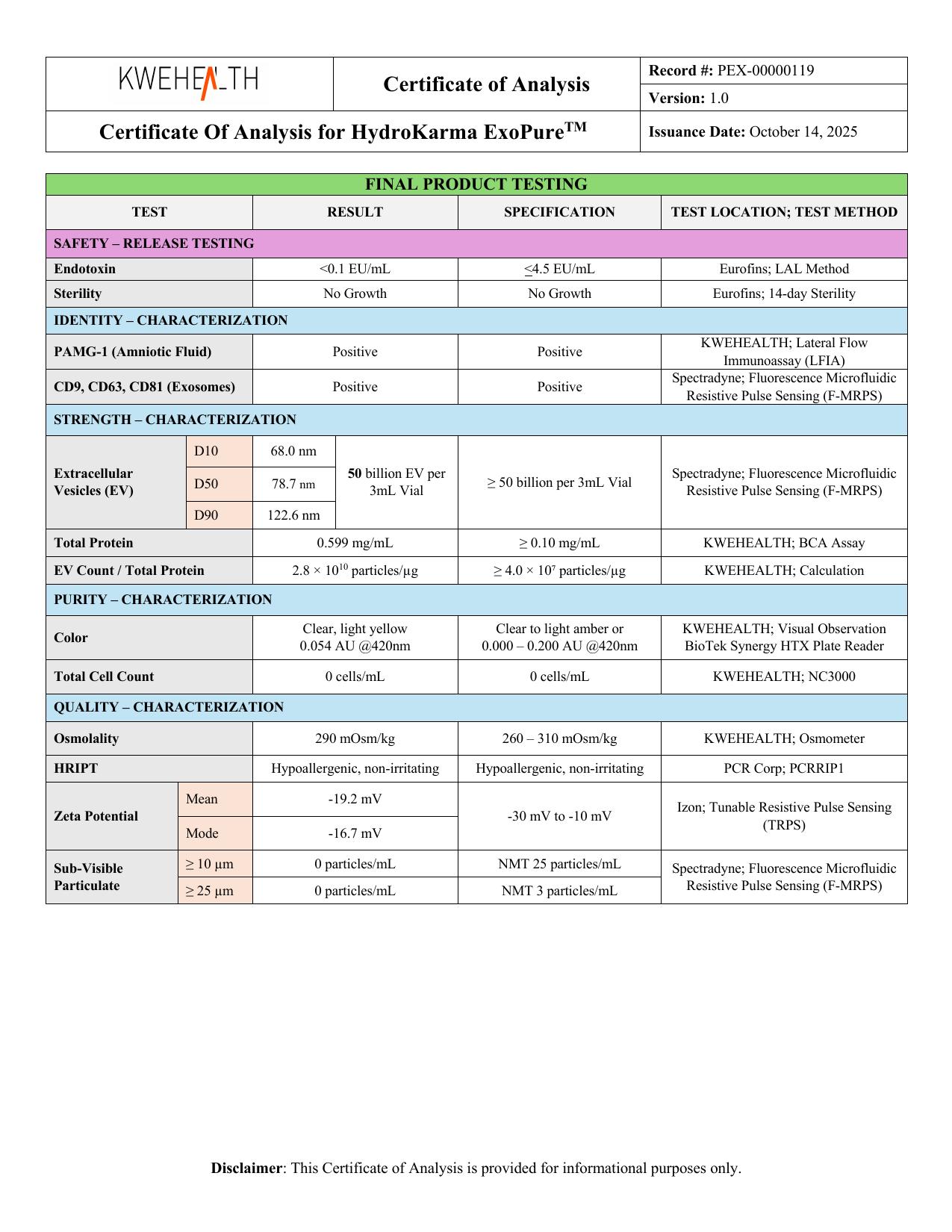
Although the proprietary purification process isn't disclosed in the COA, the finished product can still be characterized.
For HydroKarma lot KH-0007DE, KWEHEALTH reports measurements including:
- EV quantity: 50 billion EV per 3 mL vial
- Particle size: D10: 68.0 nm; D50: 78.7 nm; D90: 122.6 nm
- EV-associated markers: CD9: Positive; CD63: Positive; CD81: Positive
- Total protein: 0.599 mg/mL
- EV Count / Total Protein: 2.8 × 10¹⁰ particles/µg
- Total cell count: 0 cells/mL
These measurements characterize different aspects of the finished lot.
They do not reveal the proprietary purification workflow itself.
That is an important distinction when reading an extracellular vesicle Certificate of Analysis. For a step-by-step walkthrough, see How to Read an Exosome Certificate of Analysis (COA).
Why Doesn't the COA List Every Manufacturing Step?
A Certificate of Analysis and a manufacturing protocol serve different purposes.
A COA generally documents specified testing and results associated with a particular lot or product.
A manufacturing protocol describes how that product is produced.
Those documents shouldn't be expected to contain identical information.
A company may protect proprietary processing details while still providing batch-specific analytical results.
Therefore:
Lack of a proprietary step-by-step purification protocol on a COA doesn't mean no purification occurred.
And conversely:
A COA shouldn't be used to invent manufacturing steps that aren't actually documented.
Why the Starting Material Matters
The biological source influences the challenges involved in EV separation.
EVs isolated from cell-culture media aren't being separated from exactly the same biological environment as EVs derived from plasma, urine, milk, amniotic fluid or other biological sources.
Different starting materials contain different mixtures of proteins, lipoproteins, cells and other components.
This means a method that performs well for one source isn't automatically ideal for another.
That is another reason we should be cautious about taking a purification workflow from one scientific paper and assuming it describes how a completely different EV preparation is manufactured.
How Should You Evaluate an EV Purification Claim?
When you see claims about extracellular-vesicle purification, ask:
1. What was the starting material?
The biological source matters.
2. What separation principle was used?
Size? Density? Sedimentation? Affinity? Multiple approaches?
3. Was the method actually disclosed?
Don't assume a process based on terminology alone.
4. How was the resulting preparation characterized?
Look beyond the isolation method itself.
5. What potential non-EV components were considered?
Different starting materials present different separation challenges.
6. Are yield and purity being treated as the same thing?
They shouldn't automatically be.
7. Are conclusions supported by actual measurements?
Processing claims and characterization results should be distinguished.
These questions are much more useful than simply asking whether something was described as “purified.”
Isolation, Purification and Characterization Work Together
The complete picture looks something like this:
- Biological starting material
- Pre-processing
- Isolation / enrichment / purification
- Characterization
- Quality testing
- Lot-specific documentation
Each stage provides different information.
And the quality of an EV preparation can't be understood simply by naming one isolation technique.
Ultimately, the important question is what the complete process and resulting characterization data tell us.
The Bigger Picture
EV isolation science has evolved significantly because researchers increasingly recognize that extracellular vesicles are complex and heterogeneous.
There isn't a magical filter that lets “exosomes” through while perfectly removing everything else.
Instead, EV processing uses physical and biochemical properties to enrich selected populations.
That is why modern EV science places so much emphasis on transparent methodology and complementary characterization.
The processing method matters.
The starting material matters.
The analytical methods matter.
And the interpretation of the resulting data matters.
The Bottom Line
Extracellular vesicle isolation and purification describe processes used to separate or enrich EVs from complex biological starting materials.
Common research approaches include:
- differential centrifugation and ultracentrifugation
- density-gradient separation
- size-exclusion chromatography
- filtration and ultrafiltration
- tangential flow filtration
- precipitation
- affinity-based isolation
Each uses different physical or biochemical properties and involves its own advantages and limitations.
No single method automatically guarantees EV identity or absolute purity.
For HydroKarma products, KWEHEALTH has told Express Biologics that clinical-grade amniotic fluid is obtained through qualified tissue acquisition, delivered to KWEHEALTH, received and immediately pre-processed, and quarantined until the required test results and medical records have been reviewed—after which acceptable material is released for final manufacturing and vial filling.
KWEHEALTH also states that a subset of the finished product vials is sent out for safety testing and total exosome-count testing, and that the finished product is released for distribution once those results are acceptable.
The proprietary purification protocol itself has not been supplied to Express Biologics.
Therefore, the general EV isolation methods discussed in this article should not be interpreted as HydroKarma's manufacturing protocol.
That's ultimately one of the most important lessons when evaluating extracellular-vesicle products:
Know the difference between what scientific literature says is possible, what a manufacturer says it actually does, and what the finished-product testing actually demonstrates.
Related reading: How Are Exosomes Produced and Processed?
Original Source / References
- Welsh et al. — Minimal information for studies of extracellular vesicles (MISEV2023): From basic to advanced approaches. Journal of Extracellular Vesicles, 2024. DOI: 10.1002/jev2.12404, PMID: 38326288.
- Théry et al. — Minimal information for studies of extracellular vesicles 2018 (MISEV2018). Journal of Extracellular Vesicles, 2018. DOI: 10.1080/20013078.2018.1535750, PMID: 30637094.
- Konoshenko et al. — Isolation of Extracellular Vesicles: General Methodologies and Latest Trends. BioMed Research International, 2018. DOI: 10.1155/2018/8545347, PMID: 29662902. This review discusses major EV isolation approaches, including centrifugation, filtration, precipitation and chromatography-based techniques.
- KWEHEALTH/HydroKarma supplied manufacturing and Certificate of Analysis documentation. HydroKarma-specific sourcing, quarantine, processing, purification, characterization, vialing and lot-testing information discussed in this article comes from information supplied by KWEHEALTH/HydroKarma. The independent publications above do not describe or validate HydroKarma's proprietary manufacturing process.
Continue Learning
What Is Extracellular Vesicle Size and Why Does It Matter?
Extracellular vesicles exist across a range of nanoscale sizes. Here's what nanometer measurements mean, how D10, D50 and D90 describe a size distribution, and why size is one piece of characterization evidence rather than proof of identity.
What Is Extracellular Vesicle Purity? How EV Purity Is Evaluated
Extracellular vesicle purity is a multidimensional characterization question, not a single number on a Certificate of Analysis. Here's how particle counts, protein measurements, markers and particle-to-protein ratios each contribute — and what none of them establish alone.
Understanding Extracellular Vesicle Characterization and Testing
Characterization is how laboratories measure and describe different properties of an extracellular-vesicle preparation — particle count, particle size, EV-associated markers, total protein and zeta potential — and why no single test describes everything.